The heart is a complex organ that relies on a sophisticate electric system to function properly. Understanding the Heart Conduction System Diagram is all-important for grasp how the heart beats and pumps blood efficiently. This scheme ensures that the heart's chambers contract in a coordinated style, conserve the body's circulatory health. In this post, we will delve into the components of the heart conductivity scheme, their functions, and how they act together to keep the heart trounce.
The Components of the Heart Conduction System
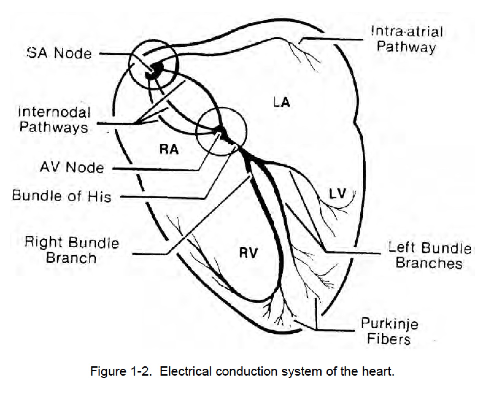
The heart conduction system consists of respective key components, each play a critical role in the heart's electric action. These components include the sinoatrial node, atrioventricular node, bundle of His, bundle branches, and Purkinje fibers. Understanding each of these parts is essential for comprehending the Heart Conduction System Diagram.
The Sinoatrial Node (SA Node)
The sinoatrial node, oftentimes name to as the heart's natural pacesetter, is locate in the right atrium. It generates electrical impulses that initiate the heart's contraction cycle. The SA node sets the heart rate, typically around 60 to 100 beats per minute in a healthy adult. These impulses spread through the atria, do them to contract and pump blood into the ventricles.
The Atrioventricular Node (AV Node)
The atrioventricular node is situated in the lower part of the right atrium, near the septum that separates the atria from the ventricles. It receives electric impulses from the SA node and delays their transmitting to the ventricles. This delay allows the atria to contract fully before the ventricles begin to pump blood. The AV node acts as a porter, see that the ventricles contract in a coordinated fashion.
The Bundle of His
The bundle of His, also known as the atrioventricular bundle, is a group of particularize muscle fibers that conduct electrical impulses from the AV node to the ventricles. It divides into the right and left bundle branches, which further distribute the impulses to the ventricular muscle fibers. The bundle of His is essential for the contemporise contraction of the ventricles, ensuring effective blood pump.
The Bundle Branches
The bundle branches are extensions of the bundle of His that impart electric impulses to the ventricular muscle fibers. The right bundle branch supplies the right ventricle, while the left bundle branch supplies the left ventricle. The left bundle branch further divides into the anterior and bum fascicles, ensuring that the left ventricle contracts uniformly. This organise condensation is all-important for preserve the heart's pumping efficiency.
The Purkinje Fibers
The Purkinje fibers are the net components of the heart conductivity scheme. They are specialized muscle fibers that find electrical impulses from the bundle branches and distribute them to the ventricular muscle fibers. Purkinje fibers ensure that the ventricles contract from the bottom up, advertise blood out of the heart and into the arteries. This organise compression is critical for maintaining blood press and circulation.
Understanding the Heart Conduction System Diagram
A Heart Conduction System Diagram provides a ocular representation of how electric impulses travel through the heart. This diagram is all-important for medical professionals and students studying cardiology, as it helps illustrate the complex interactions between the heart's components. The diagram typically includes the postdate elements:
- The sinoatrial node (SA node)
- The atrioventricular node (AV node)
- The bundle of His
- The right and left bundle branches
- The Purkinje fibers
By analyze a Heart Conduction System Diagram, one can bettor understand the succession of events that occur during a heartbeat. The diagram shows how electrical impulses rise in the SA node, travel through the atria, and are delayed at the AV node before continuing to the ventricles via the bundle of His and bundle branches. Finally, the impulses reach the Purkinje fibers, have the ventricles to contract and pump blood.
The Electrical Activity of the Heart
The electrical action of the heart can be mensurate using an electrocardiogram (ECG), which records the heart's electrical signals over time. An ECG provides worthful info about the heart's rhythm and conductivity system. The ECG waveform consists of several key components, each corresponding to a specific phase of the heart's electric activity:
- P wave: Represents the depolarization of the atria, originate by the SA node.
- QRS complex: Represents the depolarization of the ventricles, initiate by the bundle of His and bundle branches.
- T wave: Represents the repolarization of the ventricles, preparing them for the next contraction.
By analyze the ECG waveform, medical professionals can name abnormalities in the heart's conduction scheme, such as arrhythmias or conduction blocks. These abnormalities can indicate underlie heart conditions that require treatment.
Common Abnormalities in the Heart Conduction System
Several conditions can affect the heart's conductivity system, leading to unnatural heart rhythms or ineffective pumping. Some of the most common abnormalities include:
- Sinus node disfunction: A condition where the SA node fails to give electrical impulses properly, leading to an irregular heart rate.
- Atrioventricular block: A stipulation where the electrical impulses are delayed or stymy as they travel from the atria to the ventricles, often due to issues with the AV node or bundle of His.
- Bundle branch block: A condition where the electrical impulses are delayed or blocked as they travel through the bundle branches, affecting the ventricles' compression.
- Premature ventricular contractions (PVCs): Extra heartbeats that uprise in the ventricles, often due to unnatural electric action in the Purkinje fibers.
These conditions can be diagnosed using an ECG and may need treatment, such as medicine, pacemaker nidation, or other interventions, to restore normal heart map.
Diagnosing and Treating Heart Conduction System Disorders
Diagnosing heart conductivity scheme disorders involves a combination of medical history, physical examination, and diagnostic tests. The most mutual diagnostic tools include:
- Electrocardiogram (ECG): Records the heart's electrical action and identifies abnormalities in the conductivity system.
- Holter monitor: A portable device that records the heart's electrical activity over an extended period, typically 24 to 48 hours.
- Event monitor: A portable device that records the heart's electrical action when symptoms occur, providing valuable information about intermittent arrhythmias.
- Electrophysiology study (EPS): A procedure that involves inclose catheters into the heart to map its electrical action and place the source of unnatural rhythms.
Treatment for heart conduction system disorders depends on the underlie cause and rigor of the stipulation. Common treatment options include:
- Medication: Drugs that regulate heart rate, rhythm, or blood pressing, such as beta blockers, calcium channel blockers, or antiarrhythmic agents.
- Pacemaker implantation: A device that generates electrical impulses to regularize the heart rate in cases of sinus node dysfunction or AV block.
- Implantable cardioverter defibrillator (ICD): A device that monitors the heart's rhythm and delivers electrical shocks to correct life threatening arrhythmias.
- Catheter excision: A procedure that uses radiofrequency energy to destroy abnormal electrical pathways in the heart, rectify arrhythmias.
In some cases, lifestyle modifications, such as diet, exercise, and stress management, may also be commend to improve overall heart health and trim the risk of conduction system disorders.
Note: Always consult a healthcare professional for personalize advice and treatment options related to heart conductivity scheme disorders.
Preventing Heart Conduction System Disorders
While some heart conductivity scheme disorders are genetical or innate, many can be prevented or negociate through lifestyle choices and regular medical check ups. Here are some strategies to maintain a healthy heart conduction system:
- Regular exercise: Engage in regular physical action to improve cardiovascular health and cut the risk of heart disease.
- Healthy diet: Consume a equilibrize diet rich in fruits, vegetables, whole grains, and lean proteins, and low in saturated fats, sodium, and sugar.
- Maintain a healthy weight: Achieve and maintain a healthy body weight to reduce the strain on the heart.
- Avoid tobacco and limit alcohol: Quit fume and limit alcohol consumption to reduce the risk of heart disease and conductivity system disorders.
- Manage stress: Practice stress reduction techniques, such as meditation, yoga, or deep breathe exercises, to advertise overall heart health.
- Regular check ups: Schedule regular aesculapian check ups and screenings to reminder heart health and detect any abnormalities early.
By follow these healthy habits, individuals can importantly cut their risk of developing heart conductivity scheme disorders and preserve optimum heart function.
Understanding the Heart Conduction System Diagram is essential for compass the complex electric action that governs the heart s role. By larn about the components of the heart conduction system and their roles, individuals can gain a deeper appreciation for the heart s intricate workings and the importance of maintaining heart health. Regular medical check ups, a healthy lifestyle, and prompt treatment of any abnormalities can facilitate ensure that the heart conduction scheme functions optimally, upgrade overall cardiovascular health.
Related Terms:
- heart conduction scheme pronounce
- heart diagram with sa node
- bear system of heart pdf
- cardiac conductivity chart pdf
- diagram of deport system heart
- heart conduction diagram mark